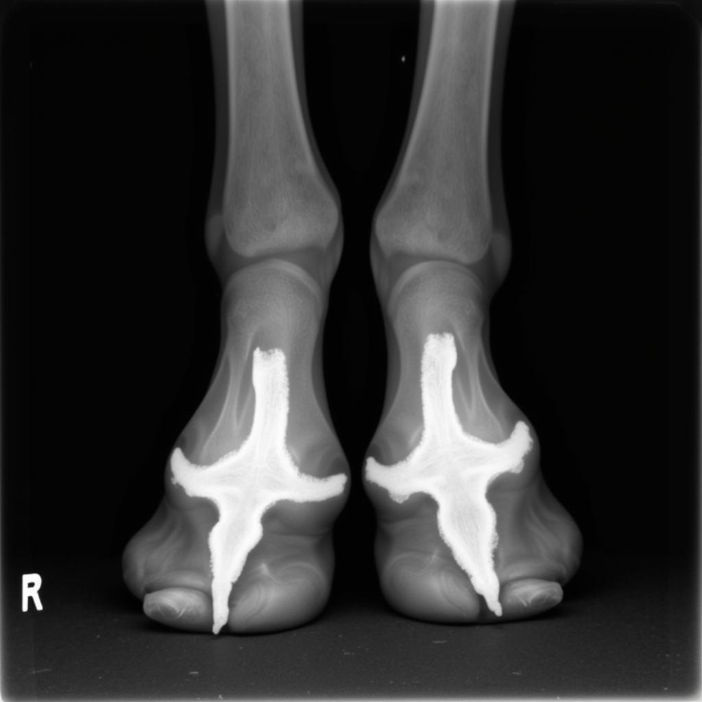
Normal Horse Hoof Radiographs are essential for diagnosing lameness and guiding farrier work. Understanding what a healthy hoof looks like on an x-ray allows veterinarians and farriers to identify deviations and develop appropriate treatment plans. This article delves into the key aspects of interpreting normal horse hoof radiographs, providing valuable insights for horse owners, farriers, and veterinary professionals alike.
Key Features of Normal Horse Hoof Radiographs
A normal horse hoof radiograph reveals a complex internal structure that must be carefully evaluated. The various components of the hoof, including the coffin bone (P3), navicular bone, and hoof wall, all have specific characteristics that indicate health. Evaluating the relationships between these structures is just as crucial as examining the individual components themselves. For instance, the angle of the coffin bone within the hoof capsule is a critical factor in hoof balance and overall soundness.
Understanding these normal features is the foundation for recognizing abnormalities. Any deviation from the normal appearance can signal a potential problem, ranging from minor inflammation to severe structural damage. Early detection through radiography can significantly improve the outcome and long-term soundness of the horse.
Positioning and Projections for Accurate Diagnosis
Different radiographic projections offer unique perspectives on the hoof’s internal structures. The most common projections include the lateromedial (side) view, the dorsopalmar/dorsoplantar (front-to-back) view, and the oblique views. Each projection highlights specific anatomical areas and is essential for a comprehensive evaluation. buck kneed in horses can sometimes be identified through careful radiographic analysis. For example, the lateromedial view is crucial for assessing the coffin bone’s angle and the relationship between the coffin bone and the hoof wall. The dorsopalmar/dorsoplantar view provides information about the symmetry of the hoof and the position of the navicular bone.
Proper positioning of the horse and the x-ray equipment is paramount for obtaining clear and accurate images. Incorrect positioning can lead to distorted images that make accurate interpretation challenging, even for experienced professionals. For example, if the hoof is not placed squarely on the x-ray plate, the coffin bone may appear to be at an abnormal angle, leading to a misdiagnosis.
Recognizing Common Variations in Normal Horse Hoof Radiographs
While there are established parameters for what constitutes a “normal” hoof radiograph, it’s important to recognize that some variations can occur naturally. Factors such as breed, age, and individual conformation can influence the appearance of the hoof on x-ray. Certain breeds, for example, might naturally have a steeper or shallower coffin bone angle than others. Recognizing these normal variations prevents misinterpretation and unnecessary interventions. horse keratoma can sometimes be confused with other hoof conditions in radiographs, highlighting the importance of a thorough evaluation.
“Understanding the range of normal variations in hoof radiographs is critical for accurate interpretation,” explains Dr. Emily Carter, DVM, a specialist in equine lameness. “It allows us to differentiate between normal anatomical variations and true pathological changes.”
How to Interpret Hoof Angles on Radiographs
The angles between different hoof structures are critical indicators of hoof balance and potential lameness issues. For example, the palmar angle of the coffin bone should be parallel to the hoof wall. Deviations from this parallel alignment can indicate imbalances that may predispose the horse to lameness.  Normal Horse Hoof Radiograph (DP View)
The Importance of Radiographs in Lameness Diagnosis
Radiographs play a vital role in the diagnosis and management of hoof-related lameness. They allow veterinarians to visualize internal structures that are not visible externally, enabling them to identify the source of pain and guide treatment decisions. horse limping is a common reason for requesting hoof radiographs. For instance, radiographs can reveal fractures, infections, and changes in bone density that may not be apparent through a physical examination alone.
“Radiographs are an invaluable tool in my practice,” states Dr. Mark Johnson, DVM, a certified farrier. “They help me pinpoint the exact location of a problem within the hoof, which is crucial for developing an effective treatment plan, whether that involves corrective shoeing, medication, or other therapeutic interventions.” shock wave horse therapy can sometimes be used in conjunction with other treatments to address certain hoof conditions.
Conclusion
Normal horse hoof radiographs are a cornerstone of equine podiatry. Understanding the key features, variations, and interpretative techniques associated with these images is essential for accurate diagnosis and effective management of hoof-related problems. By recognizing the nuances of normal hoof anatomy on radiographs, veterinarians and farriers can provide the best possible care for their equine patients, ensuring their long-term soundness and well-being.
FAQ
- What is the purpose of taking horse hoof radiographs?
- What are the different views used in horse hoof radiography?
- What are the key features to look for in a normal horse hoof radiograph?
- How can radiographs help diagnose lameness in horses?
- What are some common variations in normal horse hoof radiographs?
- How often should horse hoof radiographs be taken?
- Who can interpret horse hoof radiographs?
For any questions or assistance, contact us at Phone: 0772127271, Email: justushorses@gmail.com Or visit us at QGM2+WX2, Vị Trung, Vị Thuỷ, Hậu Giang, Vietnam. We have a 24/7 customer support team.