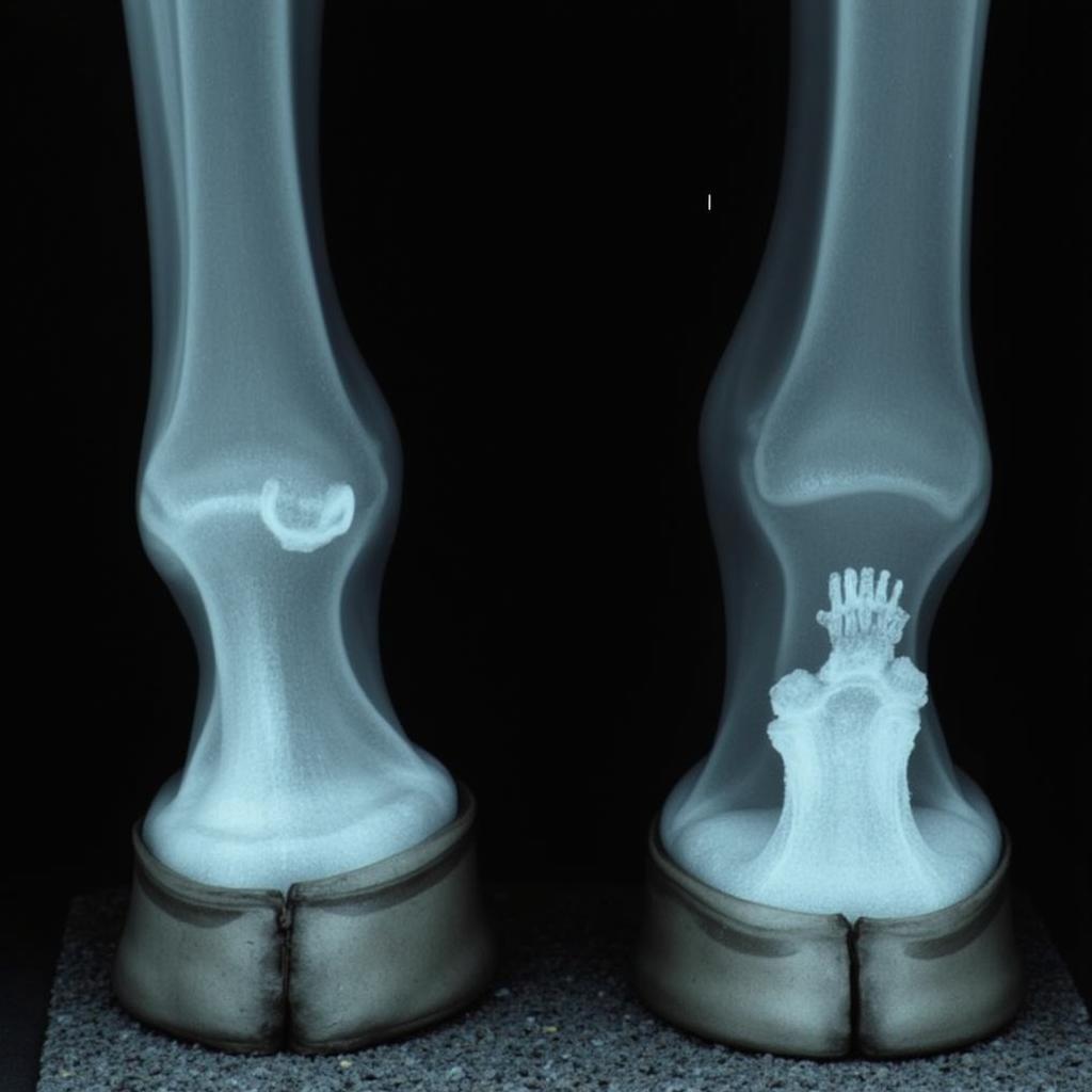
Horse foot x-rays are a crucial diagnostic tool for veterinarians, providing a detailed look inside the complex structure of the equine hoof. Understanding these images can help horse owners make informed decisions about their horse’s care, especially when lameness or other hoof problems arise.
Decoding the Mysteries of a Horse Foot X-ray
A horse foot x-ray isn’t just a picture; it’s a roadmap to understanding the intricate network of bones, tendons, and ligaments within the hoof capsule. These images allow veterinarians to identify fractures, infections, navicular disease, and other conditions that may not be visible externally. Veterinarians use specific angles and techniques to capture clear x-rays of different parts of the foot, ensuring accurate diagnosis and treatment planning. Early detection through horse foot x-rays can significantly impact a horse’s long-term soundness and overall well-being.
Why Are Horse Foot X-rays Important?
Horse foot x-rays are essential for diagnosing a wide range of conditions, from subtle changes in bone density to severe fractures. They are particularly valuable in cases of lameness where the cause is not immediately apparent. Imagine trying to fix a complex machine without seeing its internal workings – that’s what diagnosing hoof problems without x-rays would be like. These images provide a critical insight, allowing for targeted treatment and better outcomes.
“Regular horse foot x-rays, especially for performance horses, are like routine checkups for your car. They help catch small problems before they become major issues,” says Dr. Emily Carter, DVM, a renowned equine veterinarian specializing in lameness.
 Horse Foot X-ray DP View
What Can a Horse Foot X-ray Reveal?
Horse foot x-rays can reveal a multitude of issues, including:
- Fractures: X-rays can pinpoint the location and severity of fractures in any of the bones within the hoof, including the coffin bone, navicular bone, and short pastern.
- Navicular Disease: Changes in the shape and density of the navicular bone, as well as the surrounding soft tissues, can indicate navicular disease.
- Abscesses: X-rays can help locate and assess the extent of hoof abscesses, guiding treatment and drainage.
- Laminitis: X-rays can reveal changes in the laminar attachment, a key indicator of laminitis, and help monitor the progression of the disease.
- Osteoarthritis: Changes in the joint spaces and bone surfaces can suggest osteoarthritis in the distal interphalangeal joint.
symptoms of hock pain in horses
“Interpreting horse foot x-rays requires specialized training and experience,” notes Dr. David Miller, PhD, an equine radiologist. “Subtle changes in bone density or alignment can be significant indicators of underlying problems.”
Preparing for a Horse Foot X-ray
Proper preparation is vital for obtaining high-quality horse foot x-rays. This often involves cleaning and trimming the hoof to remove any debris or excess keratin that could obscure the image. In some cases, contrast agents might be injected into the hoof joint to highlight specific structures. Ensuring the horse is calm and cooperative during the procedure is also crucial.
What to Expect During the Procedure
The actual x-ray process is relatively quick. The veterinarian positions the x-ray machine and the cassette holding the x-ray film or digital sensor. They’ll then briefly activate the machine to capture the image. Several images may be taken from different angles to provide a complete picture of the hoof.
Conclusion
Horse foot x-rays are an invaluable diagnostic tool in equine podiatry. They provide crucial insights into the intricate structures of the horse’s foot, enabling veterinarians to accurately diagnose and treat a wide range of conditions. Understanding the value of horse foot x-rays can help horse owners make informed decisions regarding their horse’s health and well-being.
FAQs
- How much does a horse foot x-ray typically cost? (The cost can vary depending on the location and the number of views required. Contact your local veterinarian for specific pricing.)
- Are horse foot x-rays painful for the horse? (No, the procedure itself is not painful. However, some horses may find the positioning or the presence of the equipment slightly unsettling.)
- How long does it take to get the results of a horse foot x-ray? (If digital x-rays are used, the images are available almost immediately. With traditional film, development may take a short time.)
- Can horse foot x-rays detect all hoof problems? (While x-rays are very effective, some soft tissue injuries may not be readily visible on x-ray. Other imaging techniques, such as ultrasound or MRI, may be necessary in some cases.)
- How often should my horse have its feet x-rayed? (The frequency of x-rays depends on the individual horse’s needs. Performance horses or those with a history of hoof problems may require more frequent x-rays than those with healthy hooves.)
- What should I do if my horse is showing signs of lameness? (Contact your veterinarian immediately. Lameness can be a sign of various underlying conditions, and early diagnosis is crucial for successful treatment.)
- Can I see my horse’s x-rays? (Yes, your veterinarian will review the x-rays with you and explain the findings.)
Need help? Contact us! Phone: 0772127271, Email: [email protected] or visit us at QGM2+WX2, Vị Trung, Vị Thuỷ, Hậu Giang, Vietnam. We have a 24/7 customer support team.