Understanding the equine digestive system is crucial for any horse owner. It plays a vital role in their overall health and well-being. While many resources detail horse anatomy, an Anatomy Horse Intestine Model offers a unique, visual learning experience. These models provide an in-depth look at the complex structure of a horse’s intestines, aiding in a deeper understanding of their digestive process and potential complications.
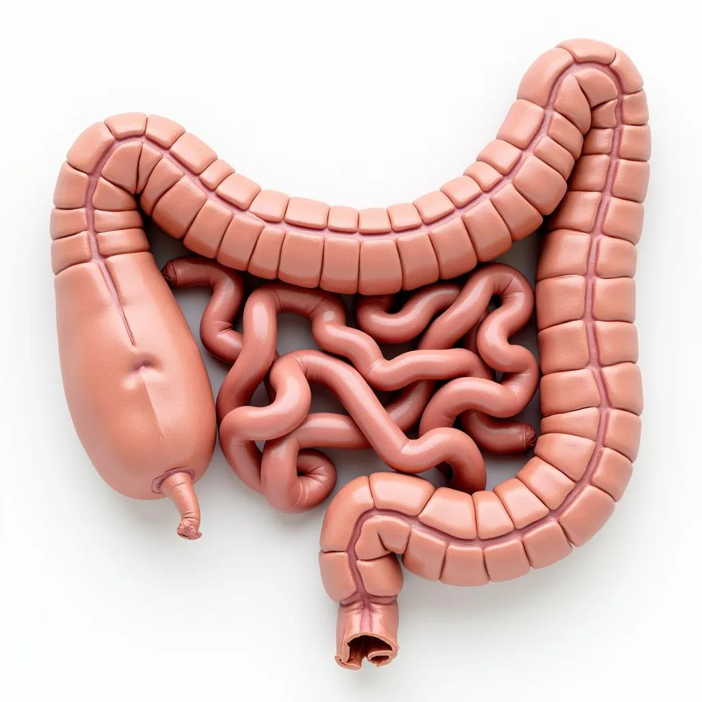 Horse Intestine Model Anatomy
Why are Horse Intestine Models Important?
Just like humans, horses rely on a healthy digestive system to absorb nutrients and eliminate waste. However, the equine digestive tract is particularly complex and susceptible to issues like colic, which can be life-threatening.
Here’s why an anatomy horse intestine model is invaluable:
- Visual Learning: Seeing a 3D representation enhances comprehension of the intricate layout of the intestines, making it easier to grasp than text or diagrams alone.
- Educational Tool: Veterinarians and educators utilize these models to illustrate digestive processes, common ailments, and treatment methods.
- Owner Understanding: Models enable horse owners to visualize how food travels through the digestive tract, emphasizing the importance of proper feeding practices and colic prevention.
Exploring the Anatomy of a Horse’s Intestine
A horse’s intestines are remarkably long, averaging around 100 feet in length! This extensive system is divided into two main sections:
1. Small Intestine:
This section, about 70 feet long, is where the initial stages of digestion and nutrient absorption occur. Key parts include:
- Duodenum: Receives partially digested food from the stomach and continues the breakdown process with enzymes.
- Jejunum: The primary site for nutrient absorption into the bloodstream.
- Ileum: Connects to the large intestine and plays a role in further nutrient and water absorption.
 Equine Small Intestine Structure
2. Large Intestine:
Comprising the remaining 30 feet, the large intestine is where fermentation takes place:
- Cecum: A large, comma-shaped pouch where beneficial bacteria break down fibrous material like hay. This fermentation process produces essential nutrients.
- Large Colon: Responsible for water absorption and formation of fecal balls.
- Small Colon: Further water absorption occurs here.
- Rectum: The final holding area for waste products before excretion.
Common Issues Visualized with an Anatomy Horse Intestine Model
These models are particularly helpful in understanding digestive complications common in horses:
- Colic: Models can demonstrate potential blockage points in the intestines, which are often the cause of colic, a serious condition requiring immediate veterinary attention.
- Parasite Infestation: Models can show how parasites can damage the intestinal lining, affecting digestion and nutrient absorption.
- Displaced Abomasum: While not directly related to the intestines, some models may illustrate the abomasum (part of the stomach) and its susceptibility to displacement, a serious condition requiring veterinary intervention.
Choosing the Right Anatomy Horse Intestine Model
Various models cater to different needs. Consider these factors:
- Purpose: Educational use for veterinary students versus owner education.
- Detail Level: Basic models showing overall structure versus those with removable parts for in-depth study.
- Material: Durable plastic for frequent handling versus more affordable, less detailed options.
“Investing in a quality anatomy horse intestine model is invaluable for any horse owner. It’s a fantastic tool to educate yourself, your staff, and your clients about the complexities of the equine digestive system.” – Dr. Emily Carter, Equine Veterinarian
Conclusion
An anatomy horse intestine model is more than just a visual aid; it’s a gateway to understanding the intricate workings of the equine digestive system. This knowledge empowers horse owners, veterinarians, and enthusiasts to provide the best possible care for these magnificent animals.
By visualizing the anatomy and common issues, we can promote better digestive health and potentially prevent life-threatening conditions. Contact us at Justus Horses USA for personalized advice on choosing the right model for your needs!
FAQs about Anatomy Horse Intestine Models:
-
Where can I purchase an anatomy horse intestine model?
You can find these models at veterinary supply stores, online retailers specializing in educational resources, and some equine tack shops. -
Are there different types of horse intestine models?
Yes, models vary in size, detail, and materials. Some are basic representations, while others offer removable parts or highlight specific digestive issues. -
How can an intestine model help me prevent colic in my horse?
By visualizing the digestive pathway, you can better understand the importance of slow diet changes, regular deworming, and providing ample fresh water – all crucial factors in colic prevention. -
Can these models be used to teach children about horse anatomy?
Absolutely! Simpler models are great educational tools for kids, fostering early interest in animal science and care. -
Are there models that show other parts of the horse’s digestive system?
Yes, some models incorporate the stomach, cecum, or even the entire digestive tract for a comprehensive view.
Do you have more questions about horse care?
Check out these related articles on Justus Horses USA:
- Understanding Equine Nutrition: A Deep Dive into Feeding Your Horse
- Colic in Horses: Signs, Symptoms, and Prevention Strategies
Need personalized advice?
Our experts at Justus Horses USA are here to help! Contact us at 0772127271 or [email protected]. You can also visit our location at QGM2+WX2, Vị Trung, Vị Thuỷ, Hậu Giang, Việt Nam. We offer 24/7 customer support to answer all your horse care questions.